The following discussion continues to build a foundation by which the reader can increase his understanding of the eletro-dermal screening process. Exposure to outside influences (interferences) impacts the body systems. In order to address these interferences, they must be detected, identified, and measured. This paper provides an overview of the evolutionary, methodical approach to develop a standard process of screening.
Process of Iteration
When I first began to research electrodermal detection, I developed a guide to locate electromagnetic skin sites according to organs and systems. I grouped the detection sites by organs and systems because most health professionals are taught to make a diagnosis using this approach.
The following list is an overview of organ and system detection sites. Each heading has many subdivisions that will be commented upon later as this journal progresses.
Organ and System Detection Sites
Allergies
Arteries
Blood
Breast
Connective Tissue
Degeneration
Ear
Endocrine
Eye
Fat Tissue
Gallbladder and Bile Ducts
Gastrointestinal Tract
Heart
Hormones
Immune System
Joints
Liver
Lymphatic System
Mucous Membranes
Musculoskeletal System
Nasopharynx
Nervous System
Odontons (Teeth)
Pancreas
Respiratory System
Skin
Spleen
Urogenital System
Veins
Palmar Arrays
Subsets of the detection sites
Each detection site has:
A name in plain language;
An electromagnetic site designation
A page for site location (bold/italic) found in the Atlas of my book, An Electrodermal Analysis of Biological Conductance
Large Intestine
Main Detection Site: LI 1b 5, 6
Greater Omentum: LI 3a 5, 6
Omental Bursa: LV 10 30
Cecum – LI 4 (R) 5
Lymph Vessels: LI 1-1 (R) 5
Superior Mesenteric (Autonomic) Plexus: SI 1a (R) 21
Smooth Muscles: LI 7 7
Appendix-Ileocecal Lymph Nodes: LI 4a (R) 5
Ascending Colon: LI 3 (R); LU5 5; 4
Lymphphatics , Cecum and Right Colon: LI 1-1 5
Superior Mesenteric (Autonomic) Plexus: SI 1a (R) 21
Peritoneum: LI 1c (R) 5
Peritoneal Lymph Vessels: LI 1b-1 (R) 5
Hepatic Flexure: LI 2 (R) 5
Superior Mesenteric (Autonomic) Plexus: SI 1a (R) 21
Sympathetic Nerve Hepatic Flexure of the Right Colon: LU 4 (R) 4
Transverse Colon, Right Side: LI 1 (R) 5
Superior Mesenteric (Autonomic) Plexus: SI 1a (R) 5
Transverse Colon, Left Side: LI 4 (L) 6
Inferior Mesenteric (Autonomic) Plexus: SI 1a (L) 21
Splenic Flexure: LI 3 (L); LU 5 (L) 6; 22
Inferior Mesenteric (Autonomic) Plexus: SI 1a (L) 21
Descending Colon: LI 2 (L) 6
Inferior Mesenteric (Autonomic) Plexus: SI 1a (L) 21
Descending Colon, Sympathetic Nerve: LU 4 (L) 4
Inferior Mesenteric Lymph Nodes: LI 4 -1 (L) 6
Sigmoid Colon: LI 1 (L) 6
Lymph Vessels: LI 1-1 (L) 6
Inferior Mesenteric (Autonomic) Plexus: SI 1a (L) 21
Inferior Mesenteric Lymph Nodes: LI 4 -1 (L) 6
Mesocolic Lymph Nodes: LI 4a (L) 6
Recto-Sigmoid Mucosa (L-3): GV 3a 61
Anus
Anal Canal: KI 5 50
Anal Sphincter: KI 4a; BL 30 50; 54
Rectum—KI 6 50
Medial and inferior (Autonomic) Plexus: KI 4 50
Venous (Hemorrhoidal) Plexus: KI 5a 50
Mucosa: SV 22 63
Recto-vesical/Recto-uterine (Douglas) Pouch: KI 6c 50
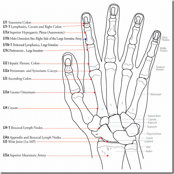
In my text An Electrodermal Analysis of Biological Conductance made an atlas demonstrating the topographical locations of these electromagnetic sites.
The graphic below is an example of one of the atlas pages.
It’s all information-codes, waves or signals
In 1996 I developed a protocol for recognizing clinical relevant non – coherent waves. The database that resulted identified the most frequent detection sites as well as the most frequent non-coherent encoded signals at those sites.
I then arranged the non-coherent codes by using plain language. I gave a short scientific description for each entry and I listed them alphabetically and by category in a glossary to be clinically useful. Were the signals related to a virus, bacteria, a fungus, toxic metal or toxic chemicals, etc.? It was my desire to make electrodermal detection a practical tool for use in the medical system as it is practiced today. Below is a list of these categories from my data.
Plain Language description of signals
Based on an analysis of 1310 files from 600 subjects from 1/8/93 to 3/8/98
Encoded Groups Number of Codes in Each Group
1. Tissue Nosodes 123
2. Bacteria 113
3. Chemicals/Drugs 112
4. Viruses 78
5. Fungi 69
6. Protozoa 53
7. Metals 46
8. Helminths 37
9. Homeopathics 30
10. Constitutionals 21
11. Cytokines 19
12. Dental 18
13. Miasms 15
14. Insects 10
15. Plants 5
16. Imponderables 5
17. Animal 4
18. Foods 3
19. Prions 3
20. Metabolites 2
Total 676
The above listing represents the broad categories of encoded non-coherent waves. There are 676 total codes in this list. Since then many more have been added.
In this section I will show how to select these waves by the process of iteration.
Iteration is defined by Merriam-Webster’s Collegiate Dictionary 10th edition as “a repetition of a sequence of operations that yields results successfully closer to the results desired”.
Recovery from illness is a biological process that takes place in a sequential fashion over time. For example, after a laceration, a clot must be formed first, followed by fibroblastic infiltration and subsequently by scar tissue formation.
In the homeopathic school of thought, Herring’s Law of Cure states that healing takes place from the inside out, from the top-down and from the acute to chronic. The analogy of peeling off the layers of an onion is often used.
Reckoning
(Merriam – Webster’s Collegiate Dictionary 10th edition 1993)
Definition: To accept something as certain; to judge
It has been found by reckoning, that electromagnetic elimination of non-coherent waves are associated with improvement in clinical conditions. The process of reckoning occurs in a sequential fashion over time.
Before we discuss the process of eliminating non coherent waves we should try to resolve the difference between a diagnosis and electrodermal signal detection
A diagnosis carries with it a precise description:
Diagnosis:
(Merriam – Webster’s Collegiate Dictionary 10th edition 1993)
1a. the art or acts of identifying a disease from its signs and symptoms
1b. the decision reached by diagnosis
2. a concise technical description of a taxon (classification).
3a. investigation or analysis of the cause or nature of a condition, situation, or problem
3b. a statement or conclusion from such an analysis
Diagnosis
(Blacks Law Dictionary, Abridged 6th edition, West Publishing Company St. Paul MN 1991)
1. A medical term, meaning the discovery of the source of a patient’s illness or a determination of the nature of his disease, from a study of its symptoms.
2. The art or act of recognizing the presence of disease from its symptoms and deciding as to its character. The decision reached for determination of type or condition through case or specimen study or conclusion arrived at through critical perception or scrutiny.
A “clinical diagnosis” is one made from the study of the symptoms only.
A “physical diagnosis” is one made by means of physical examination such as palpation and inspection.
A “ pathological diagnosis” is made by an interpretation of a pathological specimen” by a pathologist.
My definition of diagnosis is:
A consensus medical profile based on an analysis of a case history, a physical examination and an interpretation of laboratory and other technical studies for the purpose of treatment by those trained in this paradigm.
Electrodermal detection or Electrodermal Screening
Is a screening devise, in the nature of a laboratory test for gathering electromagnetic information. By itself it does not provide some of the information needed to establish a diagnosis, such as, the need for signs or symptoms or other usual laboratory data. Electrodermal detection cannot be used to make a diagnosis but the information provided can support a diagnosis. It is different from other laboratory tests in that it is an electromagnetic recognition devise, and does not provide chemical or biological information. In that sense it stands alone as a model for providing information.
The purpose of the process of iteration is, first of all, to establish an electromagnetic profile and then modify this profile in order to re-establish normal conductance.
Method of developing an electromagnetic profile
Every electromagnetic detection site on the surface of the skin represents an electromagnetic site of origin for a conducting or non-conducting electromagnetic signal.
Not all detection sites are in a convenient location for direct testing nor are all sites sufficiently sensitive to give consistent skin resistance readings by testing from that site.
When a current of injury such a pinprick, or focal pressure or electrical charge from the test probe, is applied at an inconvenient or insensitive detection site, there is amplification of the signal at that site. The injury stimulus makes the signal easier to be detected at the Main Detection Site thus making it the preferred site for applying the detecting probe.
Another way to highlight a detection site is for the one being tested to touch the detection site with the index finger of the hand holding the negative electrode; (the probe is the positive electrode.) The contacted site may then be tested at the Main Detection Site. This technique is especially useful for testing sites in “socially sensitive” areas.
Problems with detection sites:
Some detection sites are not conveniently located.
Some are on the back, others in the breast area, on the buttocks or in the groin.
Many of the inconvenient sites are also too weak to give good detection signals.
Main Detection Sites
Are located in the periphery of the fingers and toes near normal bone anatomical sites
They are easy to reach and easy to manipulate.
Are important because they represent electromagnetic events along the entire detection array especially those sites that have been highlighted or stimulated
Signals from any skin detection sites can be transferred to the Main Detection Sites of that array by a simple two-step maneuver:
1. Touch (stimulate) the desired detection site.
2. Test at the Main Detection Site of that Detection Array and observe the ohmmeter response.
Example:
If you want to determine a code at GB 5-Veins of the Head:
1. Stimulate GB 5 with the probe tip
2. Apply the probe at GB 43b, the Main Detection Site for the Gallbladder Array and observe for changes in skin resistance.
A changes at GB 43b actually reflects a changes at GB 5
Speckhart’s Protocol for Electromagnetic Profile Development
Purpose of the Protocol: To rapidly survey detection sites and codes.
The following is a list of some the more frequently used detection sites.
H-H Hand to Hand
Main Detection Sites
LY 1-2 Head and Neck
LU 10c Lungs
LI 1b Large Intestine
NV 1b Peripheral and Central Nervous System
CI 8d Circulation (Arteries, Veins, Lymphatics)
AL 1b Allergies
OR 1b Cellular Metabolism
TW 1b Endocrine System
HT 8c Heart
SI 1b Small Intestine
Sp 1a (R) Pancreas
Sp 1a (L) Spleen
AR 1b Joints
ST 44b Stomach
FI 1b Connective Tissue
SK 1-3 Skin
FA 1b Fatty Tissue
GB 43b Gallbladder and Bile Ducts
KI 1-3 Kidney
BL 66b Urinary Bladder and Urogenital Organs
Other Important Detection Sites
TW 20 Hypothalamus
OR 1-1 Lymphatics of Organ Degeneration
LY 3 Nose and Paranasal Sinuses
LY 2 Jaw and Teeth
SV 48 Striated Muscle
GB 17 Reticular Formation
SV 61 Eosinophils
GB 5 Veins of the Head
SV 46 Periosteum
AR 1c Synovial Membranes
NV 1c Spinal Cord and Meninges
